
2D Echocardiography (also known as a 2D Echo) is a type of ultrasound test that uses sound waves to create images of the heart. It’s a non-invasive, widely used diagnostic tool that helps doctors assess the heart’s structure, function, and overall health.
How 2D Echocardiography Works:
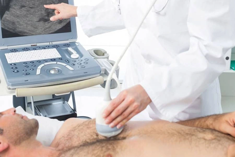
- Sound Waves:
- A small device called a transducer is placed on your chest. This device emits high-frequency sound waves (ultrasound) that bounce off the heart’s structures, like the heart valves and chambers.
- The reflected sound waves are then converted into images that appear on a monitor.
- Creating Images:
- Unlike traditional X-rays, 2D echocardiography produces real-time, moving images of the heart. These images can show the heart’s chambers, valves, blood flow, and even the heart muscle’s movement.
- No Radiation:
- 2D echocardiography doesn’t involve radiation, making it a safer option for repeated testing.
What Does 2D Echocardiography Show?
2D Echocardiography is primarily used to assess the following:
- Heart Chambers and Valves:
- It shows the size, shape, and motion of the heart’s four chambers (the left and right atria and ventricles) and the heart valves (mitral, tricuspid, aortic, and pulmonary valves). It helps to evaluate if the valves are working correctly and whether there are any abnormalities such as leakage (regurgitation) or narrowing (stenosis).
- Heart Muscle Function:
- It can evaluate how well the heart is pumping blood. The movement of the heart muscle can be assessed to check for signs of heart failure or damage caused by heart attacks.
- Blood Flow:
- By using Doppler ultrasound along with 2D echo, it shows how blood flows through the heart’s chambers, valves, and vessels. This can help detect issues like blood clots, abnormal flow patterns, or narrowing of the heart’s blood vessels (coronary artery disease).
- Heart Wall Motion:
- The test helps assess if all areas of the heart are moving normally. Abnormalities in wall motion may indicate previous heart attacks, heart muscle disease (cardiomyopathy), or other conditions.
How 2D Echocardiography Is Performed:
- Preparation:
- Typically, no special preparation is required. You may be asked to avoid eating or drinking for a few hours before the test.
- You’ll be asked to lie on your back on an examination table.
- Electrode Placement:
- Electrodes are placed on your chest to monitor your heart’s electrical activity during the procedure (similar to an ECG).
- The Procedure:
- A gel is applied to your chest to help the transducer make good contact with your skin.
- The technician or cardiologist moves the transducer over your chest to capture various images of your heart from different angles. You may be asked to change positions or hold your breath briefly to get clearer images.
- Duration:
- The test usually takes about 30 minutes to an hour.
Why 2D Echocardiography Is Done:
2D echocardiography is used for a wide range of heart-related issues, such as:
- Heart Murmurs: To investigate the cause of an abnormal heart sound, often due to valve problems.
- Chest Pain: To determine whether chest pain is due to heart disease or another cause.
- Shortness of Breath: To assess if heart failure or another heart condition is the cause.
- Heart Failure: To evaluate the pumping function of the heart in cases of heart failure.
- Heart Valve Disease: To detect abnormalities like valve narrowing, leakage, or prolapse.
- Infections or Inflammation: To check for conditions like endocarditis (infection of the heart valves).
- Post-Heart Attack: To evaluate heart function after a heart attack or coronary bypass surgery.
Advantages of 2D Echocardiography:
- Non-invasive: There are no incisions, injections, or radiation involved.
- Safe: No risks associated with the procedure, making it suitable for almost all patients.
- Real-Time Results: Provides immediate images of the heart, helping doctors make quick decisions.
- Comprehensive: Allows doctors to examine the structure, function, and blood flow of the heart.
Limitations:
- Image Quality: In some cases, the quality of the images may be limited due to factors like body type (e.g., obesity), lung interference, or patient movement.
- Requires Expertise: The interpretation of the results requires a skilled echocardiographer and cardiologist.