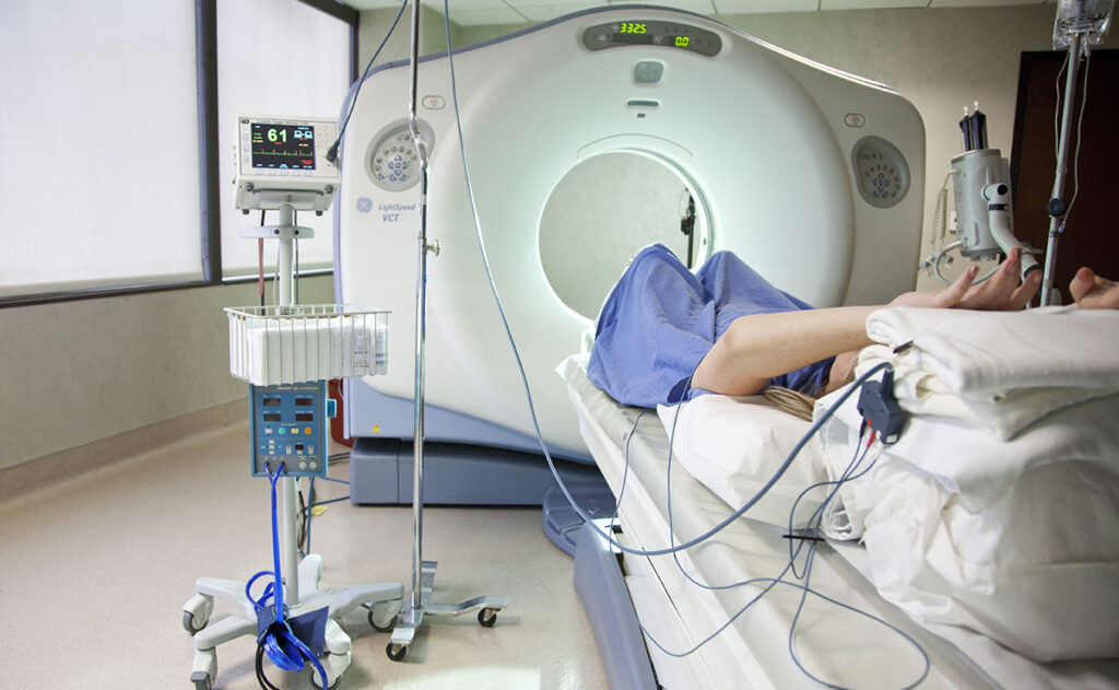
Angiography is a medical imaging technique used to visualize the inside of blood vessels and organs, most commonly the heart, brain, or kidneys. It’s a valuable test for diagnosing issues like blockages, aneurysms, or other vascular conditions. There are different types of angiography, with coronary angiography being the most common when it comes to heart health.
What is Coronary Angiography?
Coronary angiography involves injecting a special dye (contrast agent) into the coronary arteries through a catheter inserted into a blood vessel, usually in the groin or wrist. This dye allows the arteries to be seen on X-ray images, revealing any blockages, narrowing, or other abnormalities in the blood vessels supplying the heart.
How is Angiography Performed?
The procedure is minimally invasive and usually done in a hospital’s cardiac catheterization lab (also called a cath lab). Here’s what typically happens during coronary angiography:
- Preparation:
- You may be asked to avoid eating or drinking for a few hours before the procedure.
- An intravenous (IV) line may be inserted into your arm for medications or fluids.
- You may receive a mild sedative to help you relax, but you’ll be awake during the procedure.
- Catheter Insertion:
- A small incision is made in the groin or wrist area, and a thin tube (catheter) is inserted into a blood vessel. The catheter is carefully guided toward the coronary arteries using real-time imaging, such as fluoroscopy.
- Injection of Contrast Dye:
- Once the catheter reaches the coronary arteries, the doctor injects a special dye (contrast agent) into the arteries.
- X-rays or fluoroscopy are used to take detailed images of the coronary arteries as the dye flows through them, showing any blockages or abnormalities.
- Assessment:
- The images generated help doctors identify any narrowed or blocked areas in the coronary arteries. They can also assess the severity of the blockages.
- Procedure Completion:
- The catheter is removed, and pressure is applied to the insertion site to prevent bleeding.
- The entire procedure usually takes around 30 minutes to an hour.
Why is Coronary Angiography Performed?
Coronary angiography is often performed when a doctor suspects a blockage or narrowing of the coronary arteries that may be causing heart problems. Some common reasons for the procedure include:
- Chest pain (Angina): To determine if blocked or narrowed coronary arteries are causing chest pain.
- Heart attack: After a heart attack, angiography is often done to evaluate the extent of the damage and identify any blockages that need treatment.
- Unexplained shortness of breath: Angiography can help assess if heart disease or coronary artery issues are contributing to breathing difficulties.
- Risk of heart attack: In people with high risk of heart disease, angiography can be used to assess the presence and severity of coronary artery disease.
- Assessment after heart surgery or stent placement: Doctors may perform angiography to monitor how well stents or bypass grafts are functioning.
Risks and Complications
While coronary angiography is generally safe, it carries some risks, including:
- Bleeding or infection at the catheter insertion site.
- Allergic reaction to the contrast dye used in the procedure.
- Kidney problems: The contrast dye can sometimes affect kidney function, especially in patients with pre-existing kidney conditions.
- Heart attack: Although rare, there is a risk of causing a heart attack or damaging the heart during the procedure.
- Arrhythmias: The procedure may occasionally cause irregular heart rhythms.
- Stroke: Very rarely, a stroke can occur during angiography due to the catheter or clot formation.
What Happens After Angiography?
- Recovery: After the procedure, you’ll be monitored in a recovery room for a few hours. If the catheter was inserted through the groin, you may need to lie flat for several hours to avoid bleeding from the insertion site.
- Discharge: Most people can go home the same day or the next day, depending on how well they recover.
- Post-procedure care: You may be given specific instructions on how to care for the catheter insertion site, activity restrictions, and follow-up appointments.
Treatment Options Following Angiography
If a blockage or narrowing is found, there are several potential treatments that may be considered:
- Angioplasty and Stent Placement:
- If significant blockages are found, the doctor may perform angioplasty (a procedure to widen the artery) and place a stent (a small mesh tube) to keep the artery open. This is often done immediately after the angiogram.
- Coronary Artery Bypass Surgery (CABG):
- In some cases, if the blockages are too severe or widespread for angioplasty and stenting, bypass surgery may be recommended. This involves creating new pathways for blood flow around the blocked arteries.
- Medications:
- Medications like blood thinners, statins, or beta-blockers may be prescribed to manage cholesterol, prevent blood clots, or control blood pressure.
- Lifestyle Changes:
- After an angiogram, it’s crucial to adopt a heart-healthy lifestyle, including eating a balanced diet, exercising regularly, managing stress, and avoiding smoking.
Alternative Tests to Angiography
In some cases, doctors may use other non-invasive imaging tests to assess heart function and blood vessels, such as:
- CT Angiography (CTA): A less invasive alternative that uses a CT scan and contrast dye to visualize blood vessels.
- Stress Tests: A test where the heart is monitored while exercising or through medication to simulate exercise, assessing how the heart reacts to stress.
- Magnetic Resonance Angiography (MRA): Uses magnetic fields and radio waves to create detailed images of blood vessels.


