ARRHYTHMIA
Causes
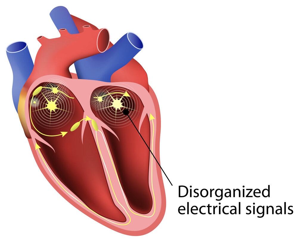
Arrhythmias occur if there is a block or delay in the electrical pathways, irregular signal generation from pacemaker node, irregular impulse conduction or due to impulse generation in any part of heart except the pacemaker node. This could happen:
- As a side effect of excessive smoking, alcohol use, caffeine and nicotine use, illicit drug abuse, or as side effect of certain over the counter or prescription drugs.
- Due to strong emotional stress and anger outbursts.
- Due to high blood pressure, coronary heart disease, rheumatic heart disease, heart attack, heart failure, or under or over active thyroid gland.
- Since birth called congenital arrhythmia (for example Wolff-Parkinson-White syndrome).
- At times no cause is found.
Types of Arrhythmia
The four main types of arrhythmia are extra or premature beats, supraventricular arrhythmias, ventricular arrhythmias, and bradyarrhythmias.
- Premature BeatsThese are the most common types of arrhythmia. They are of two types depending on where the extra betas occur: premature atrial contractions (PAC) and premature ventricular contractions (PVC).Both PAC and PVC are usually harmless, can occur without a cause or due to stress, exercise, caffeine or nicotine intake, and need no treatment. There are no signs and symptoms except a feeling of flutter in chest or extra heart beat. At times they occur in heart disease and need treatment.
- Supraventricular ArrhythmiasSupraventricular arrhythmias are basically tachycardias or tachyarrythmias originating in the atria or AV node. Examples: atrial fibrillation, atrial flutter, and paroxysmal supraventricular tachycardia (PSVT), congenital (Wolff-Parkinson-White syndrome)
- Atrial FibrillationIn atrial fibrillation electrical impulse is generated anywhere in the atria or pulmonary veins and travels through out the atria in a disorganized manner. The atria quiver or fibrillate in a fast and irregular way instead of contracting properly.Cause: Usually occurs due to coronary heart disease, rheumatic heart disease or high blood pressure, in hyperactive thyroid gland, with advanced age, or in alcohol abuse. Sometimes no cause is identified.Complications: When rate of quivering becomes as 300 per minute or more, it may cause quivering of ventricles and heart failure. Blood pools in quivering atria and forms clots, which can break and travel to brain causing stroke.
- Atrial Flutter
This is similar to atrial fibrillation, except that the electrical impulses travel in an organized manner. - Paroxysmal Supraventricular TachycardiaPSVT occurs when the signals go from atria to ventricles and come back to atria causing extra beats and short spurts of very fast heart rates. PSVT can occur during physical activity or without a cause in young people. Usually it is not dangerous.In another PSVT type in Wolff-Parkinson-White syndrome, electrical impulses travel from the atria into the ventricles along an extra pathway as well.
- Ventricular ArrhythmiasVentricular arrhythmia start in the ventricles, are usually serious and require urgent medical care. They occur in heart attack, coronary heart disease, and conditions causing weak heart muscles.
There are two main types of ventricular arrhythmias: - Ventricular TachycardiaVentricular tachycardia is spurts of very fast but regular beating of ventricles. It is the less serious type of ventricular arrhythmia, and can become dangerous only if it lasts long.
- Ventricular Fibrillation (V-fib)In V-fib, highly disorganized electrical signals make the ventricles quiver or fibrillate instead of contracting properly. This can be extremely dangerous as it can result in sudden cardiac arrest and death. This needs urgent treatment by electrical shock therapy through a defibrillator.In addition to above mentioned causes, it can occur if you suffer from potassium, calcium, and magnesium imbalance due to kidney disease or as effect of certain medications.
- BradyarrhythmiasBradyarrhythmias are heart rates slower than 60 beats per minute. This can be a normal finding in active adults. However, bradyarrhythmia can occur due to electrolyte imbalance, heart attack, underactive thyroid or use of drugs such as beta blocksrs, digoxin, calcium channel blockers.
Symptoms
Arrhythmias can occur without signs and symptoms or cause mild ones such as:
- Palpitations (sensing fast or irregular heart beats)
- Irregular heartbeat
- Slow heartbeat
- Feeling as though heart pauses in between beats
Serious symptoms of arrhythmia include:
- Anxiety
- Shortness of breath
- Sweating
- Chest pain
- Dizziness, light-headedness, fainting or fainting like sensation
- Feeling weak
Diagnosis
It is often difficult to diagnose arrhythmias as their symptoms occur once in a while and may be difficult to detect. Your doctor can diagnose arrhythmia by taking your medical and family history, doing a physical examination and certain tests.
Medical and Family Histories
Your doctor would:
- Note your symptoms
- Ask you regarding other health problems you or your family member may have such as high blood pressure, heart disease, diabetes, thyroid disorders etc.
- Note the medications you are taking including any over the counter medications or supplements.
- Ask you about your habits such as smoking, alcohol use, illicit drug use, level of physical activity, and whether you have physical or emotional stress and are prone to anger.
- Ask whether someone in your family was ever diagnosed with arrhythmia or died suddenly.
Physical Examination
During your physical examination, your doctor may:
- Listen to your heart and lung sounds
- Hear an abnormal or unusual heart sound called a murmur.
- Check your pulse
- Check your legs and feet for swelling
- Check in general for other diseases such as thyroid swelling
Diagnostic Tests and Procedures
Main Tests
- Electrocardiogram (EKG/ECG): This simple test helps record your heart beat. It tells the doctor about irregular heart beats, fast beats, previous heart attacks, and problem in the conduction of impulses in the heart.
- Holter and Event Monitors: Sometimes the ECG taken over a few seconds or minutes is not able to detect an arrhythmia. Hence you may be given a holter or event monitor to wear. These are small machines with leads or electrodes will be attached to your chest to record your heart beat. The recorder can be kept in a pocket or slung over your neck. You have to wear it for 24-48 hours while you go about your normal routine.Holter can take continuous recording of your heart for 24-48 hours while event monitor does this at certain times. Both can be worn for a week also.Both machines may have a push button which you can press if you have symptoms. When you have symptoms, you may also be able to send the electrical signals of your heart to a central database.
Other Tests
Your doctor may or may not opt for these tests depending on your symptoms.
- Blood tests: Your blood may be drawn to test for electrolytes (such as potassium, calcium, magnesium), blood sugar, or thyroid hormones.
- Chest X Ray: This can show a large heart or fluid in lungs . It can also tell the doctor if you have a lung disease.
- Echocardiography (ECHO): This is like an ultrasound of heart where your doctor can see your moving heart. This test tells your doctor about the size of heart and its chambers, the thickness of the walls of the artery, contraction of heart, mixing of blood, previous heart attack, force of blood flow etc.
- Stress Test: If you are capable of running on a tread mill, your doctor may take you up for this to examine how your heart behaves under stress. An ECG recording and blood pressure monitoring will be done while you run. The test is often followed up with ECHO and nuclear heart scans.
- Coronary Angiography: Your doctor may ask for this test if he suspects a block in the heart arteries. A dye is injected through a tube placed in the blood vessel of your groin or arm and taken up to your heart. The doctor can see the flow of the dye on a screen in front of him.
- Electrophysiology study (EPS): In this test a thin wire will be inserted through a vein in your upper thigh or arm and guided to the heart to record the electrical signals. The doctor can electrically stimulate your heart to see if arrhythmias are occurring or not. The doctor can also record the signals after giving an anti-arrhythmia medication to see if it is working or not.
- Tilt table testing: If you complain of fainting spells, your doctor may request this test to rule out other causes of fainting. You will lie on a table that will move from horizontal to upright position to see if you faint. While the table moves, your doctor will record your ECG, heart rate, blood pressure and symptoms.
- Implantable loop recorder: This device is placed under the skin of the chest by a minor surgery. This helps detect symptoms and electrical abnormality over a period of 12 to 24 months in patients who rarely have symptoms.
Treatment
Arrhythmias which cause symptoms and are serious can be treated with medication, medical devices and procedures, surgery and Vagal maneuvers. Serious arrhythmias are those which are likely to cause complications like stroke, heart failure and sudden cardiac arrest.
Medicines
Medications used to treat arrhythmias are called antiarrhythmics. Some of these medications have serious side effects. You must take the medications as prescribed. Ask your doctor about their potential side effects. You must also immediately report any new symptom or recurrence of an old symptom.
Antiarrhythmic medications can:
- Sow down a fast rate: Examples: beta blockers, calcium channel blockers (such as diltiazem and verapami, and digoxin.
- Restore the normal rhythm of heart: Examples: amiodarone, disopyramide, sotalol, procainamide, quinidine, etc.
Apart from antiarrhythmics, your doctor may prescribe blood thinners or medicines to control thyroid disorders.
Medical Devices and Procedures
- Pacemaker: Your doctor will place this small device under the skin of your chest or abdomen to control abnormal heart rhythms. The device senses an abnormal heart rhythm and sends out electrical signals to normalize the rhythm. Also, used to treat bradyarrhythmias.
- Cardioversion: In defibrillation or cardioversion, a device called defibrillator is used to give jolts of electricity to the body to treat arrhythmia. Sometimes a defibrillator device is placed under the skin of the chest to treat ventricular arrhythmias. This device is called implantable cardioverter defibrillator (ICD), whichsends out electrical shocks when it senses abnormal rhythms.
- Catheter Ablation: As in EPS study, a wire is inserted through a vein in your upper thigh or arm and guided to the heart, anda special machine is used to send energy through this wire to destroy small portions of heart tissue which are generating the abnormal rhythm.
Surgery
- Maze Surgery: Sometimes during other heart surgeries such as surgery for repair of a heart valve, your doctor may perform another surgery to correct your arrhythmia. This is called maze surgery. Small cuts or burns are given to the atria to prevent spread of abnormal electrical signals.
- Coronary artery bypass surgery is done to if coronary heart disease is the cause of arrhythmia. This surgery improves blood flow to the heart and prevents arrhythmia.
Vagal maneuvers
Simple exercises can slow down or stop abnormal heart rhythms by affecting the Vagus nerve. However, please do not do these exercises without consulting your doctor.
Vagal maneuvers which can help are:
- Coughing
- Gagging
- Gently pressing eyelids with fingers
- Splashing ice cold water on face

